The team in ED need to understand the rationale for the brain imaging they are requesting. Drag the words to describe the rationale for his imaging.
Iain returns from CT and the medical team review the imaging with the neuro radiologist.
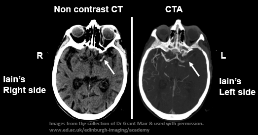
CT scan showing early infarct on left side and CTA showing blocked left MCA.
Brain scans by Grant Mair is licensed under CC BY-NC-SA 4.0
Iain’s non-contrast CT scan excludes a bleed but shows a dense (white) middle cerebral artery (MCA) on the left due to clot within the artery. There are also some early ischaemic changes (loss of grey/white matter discrimination) visible. A blocked proximal MCA is confirmed on the CTA (arrow).
Drag the words
Iain required an immediate *non-contrast* CT brain scan to exclude a cerebral *bleed* due to him taking an *anticoagulant*. Although, the onset is still less than an hour ago he is not *eligible* for treatment with *thrombectomy* because he is taking *apixaban*. *alteplase* is still an option, so they request a *CTA* to establish whether he has a blocked large *artery*.