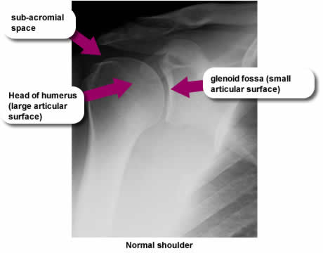
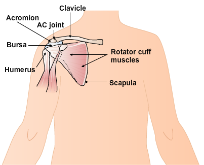
The integrity of the shoulder joint relies on muscular strength because as you can see left, the bony articular surfaces of the head of humerus and glenoid fossa differ in size to allow for a large range of movement at the shoulder. This results in a natural instability which means that the shoulder is at risk of dislocation and damage. Control of the range of motion at the shoulder involves a number of small muscles whose tendons pass between the moving bony surfaces. These may easily be damaged if the bony alignment is compromised.
Investigations
 X-ray
X-ray
Useful in identifying bony displacement and / or underlying fractures. A thorough clinical assessment will inform the best physical management, however in the presence of hemiplegia, precisely locating the anatomical structure involved is difficult as many of the diagnostic tests cannot be used. The presence or absence of subluxation can be determined by using palpation of the sub-acromial space.
MRI
Scanning has been used for diagnostic purposes but all imaging must be used with caution as similar pathological changes may be seen in people of a similar age who do not report pain.
Ultrasound
In the chronic intractable stages of shoulder pain ultrasound may be used to identify structures which may then be treated by injection with local corticosteroid.
Page last reviewed: 18 Feb 2020