- Your visual field is the visual information that is seen (both centrally and peripherally) when you look straight ahead.
- Visual field loss that occurs following a stroke commonly affects the vision of both eyes and the area of sight loss is always contra-lateral to the stroke location.
- It occurs equally often in right and left hemisphere strokes.
- Visual field loss can be one of the presenting symptoms of stroke, often described by the person as blurred or obscured vision. People may complain of visual field loss in one eye and explain that their vision ‘is not right’.
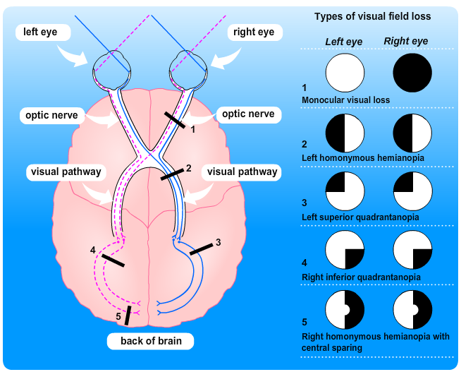
Visual field loss can occur in many different forms and the diagram below illustrates the visual pathway and the potential defects. A stroke that causes visual field loss can affect the pathway at 2, 3, 4 or 5. Strokes that only affect the visual fields most commonly affect the occipital lobe – position 5. Strokes which cause a combination of problems such as unilateral limb weakness, speech problems AND visual field loss more often affect positions 2, 3 and 4. Although not classified as strokes (because they don’t affect the brain itself) ischaemic lesions in the optic nerve (position 1) or retina can lead to loss of vision in one eye only. Traditionally, transient loss of vision in one eye due to an ischaemic lesion is referred to as amaurosis fugax and is classified rather confusingly as a TIA.
For a short explanation of each of the types of visual field loss illustrated in the drawing above, see the Additional Information box below.
As you will have seen from the hierarchy of vision shown in the module introduction (see Additional Information box to view this again), visual fields form part of the base of the pyramid. They provide us with full and clear visual information upon which the higher levels of visual processing depend.
For more information about screening, assessment and management of vision problems in the first 30 days after an acute stroke see the following best practice statement: Best Practice Statement for Screening, Assessment and Management of Vision Problems in the First 30 Days after an Acute Stroke
The diagram illustrates visual defects caused by stroke with a horizontal cross-section of the brain, optic nerves and eyes, as viewed from above.
Monocular visual loss
Occurs at the right optic nerve and affects the right eye in this example, but not the left.
Left homonymous hemianopia
Occurs along the right-sided visual pathway, and obscures the left side of vision in both eyes.
Left superior quadrantanopia
Occurs in the right side of the brain, and affects the upper left field of vision in both eyes.
Right inferior quadrantanopia
Occurs in the left side of the brain, and affects the lower right field of vision in both eyes.
Right homonymous hemianopia with central sparing
Occurs in the left side of the back of the brain, and affects the whole right field of vision in both eyes, except for the centre of the field of view.

Page last reviewed: 29 Jul 2021