Bill attends the x-ray department for his videofluoroscopy and is given a small amount of food and fluid of different consistencies, mixed with a contrast material such as barium. The moving image allows the SLT to track the pathway of the food/fluid through the oral cavity and pharynx and detect any signs of a swallowing impairment at these levels.
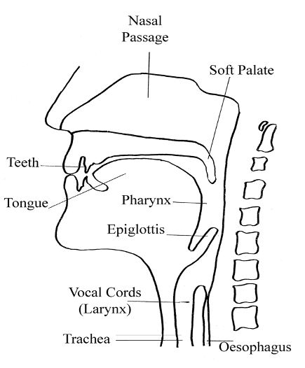
The diagram below shows the anatomical structures required for swallowing.
 |
Video by kind permission of Nutricia.
(This video contains sound, please ensure that the computer you are using has sound enabled and speakers or headphones. You can enlarge the video by clicking on the enlarge icon in the control bar under the video clip. Please note: videos may not load if you are accessing these through an NHS firewall.)
Bill has difficulty clearing his pharynx. The pharynx is the back of your throat. It sits behind the mouth, below the nose and above the trachea (airway) and oesophagus (gullet).
Can you spot the moment when Bill aspirates? A person should cough to expel the material from their airway but a cough reflex may be delayed or absent after a stroke. If the cough reflex is not triggered, this is known as silent aspiration.
The outcome of his videofluoroscopy shows that Bill is unsafe to swallow all oral diet and fluid consistencies at this time. The MDT, in discussion with Bill and his family, decide as a first step to pass an NG tube. The Speech and Language Therapist will continue to review Bill’s swallowing status and devise a swallowing therapy programme for him.
Side-on diagram shows human mouth and throat, detailing features relevant to eating and swallowing, showing location of teeth, tongue, nasal passage, soft palate, pharynx, epiglottis, vocal chords (larynx), trachea, and oesophagus.
Video script: Videofluoroscopy is a form of live action x-ray that allows the speech and language therapist to examine a patient’s swallow.
Page last reviewed: 03 Jun 2021