Symptoms & triggers

It is important to note that an individual with inherited LQTS may be asymptomatic until a QT interval prolonging drug is taken. Margaret had no cardiac symptoms until she commenced trimethoprin. Following her aborted sudden cardiac death episode, she was admitted to the Coronary Care Unit for observation and further investigation.

Please enable JavaScript in your browser to see this interactive content.

Pulse point

The fact that Margaret has a family history of Sudden Cardiac Death should be a red flag trigger to consider inherited LQTS.


Symptoms Triggers
  • Syncope (often misdiagnosed as common faint or seizure)
  • Palpitations
  • Sudden death
  • Sudden loss of consciousness during physical exertion or during emotional excitement should strongly raise the possibility of LQTS
  • Family history of unexplained syncope or sudden death in young people should also raise suspicion of LQTS
  • QT interval prolonging medications
  • Swimming, running
  • Startle: alarm clock, loud horn, ringing phone
  • Emotions: anger, crying, test taking or other stressful situations
  • Sudden death may also occur during sleep
  • Electrolyte imbalance

Drug induced LQTS

Several medications, including drugs prescribed for non-cardiac indications, have been associated with a prolongation of the QT interval on the ECG. Under certain circumstances, including patients with congenital LQTS, these drugs will excessively prolong the QT interval, triggering polymorphic ventricular tachycardia. Drugs that prolong the QT interval belong to several pharmacological classes, but most of them share one pharmacological effect: they lengthen cardiac repolarization, primarily by blocking specific cardiac K (Potassium) channels. The potent blocking of cardiac K channels and excessive lengthening of cardiac repolarization leads to prolonged QT intervals.

Patients with QT prolongation on ECG or a genetic predisposition to LQTS should avoid QT prolonging drugs. These drugs will prolong QT even in a normal person. They include drugs available on prescription and those that may be purchased over the counter.

Please enable JavaScript in your browser to see this interactive content.

Margaret has been taking trimethoprin, as prescribed, for chronic urinary tract infections. Trimethorpin is known to prolong the QT interval. This is discontinued on admission to hospital.

What is inherited Long QT Syndrome?

Given her clinical presentation, medical staff suspect that Margaret may have inherited Long QT Syndrome (LQTS). QT is a measure of the time between the start of the Q wave and the end of the T wave in the electrical cyle of the heart. LQTS is diagnosed when the QT measures more than/= 480mn in repeated 12 lead ECGs. (In a patient with unexplained syncope, if the QT measures more than/=460 in repeated 12 lead ECGS, then LQTS should be considered.)

LQTS causes an abnormality of the heart’s electrophysiology. While the mechanical function of the heart is entirely normal, there is an electricophysiological problem due to defects in the ion channels of the heart muscle cells. Ion Channels are important for the electrical activation (depolarisation) of the heart that results in the cardiac contraction and electrical recovery (repolarisation).

Please enable JavaScript in your browser to see this interactive content.

These ion channel defects cause QT prolongation and predispose to a very fast and malignant heart rhythm called Torsades de Pointes which, if sustained, may lead to sudden loss of consciousness (syncope) and sudden cardiac death.

Inherited LQTS is the most common channelopathy and occurs in approximately 1:2000 people.

In most cases of LQTS (LQTS 1, LQTS 2), the potassium channels that regulate the movement of potassium ions from the inside to the outside of the cells are affected. In a smaller proportion of cases (LQTS 3), a sodium channel that regulates the flow of sodium from the outside to the inside of cells is affected.

Inherited: Case 1: Margaret

Margaret portrait

Meet Margaret

Margaret in hospital bed, her daughter visiting

Margaret is a 65 year old lady with diabetes mellitus. She suffers from chronic urinary tract infections and her GP has prescribed trimethoprin. When Margaret collapsed at home, her daughter successfully performed cardiopulmonary resuscitation until paramedics arrived and re-established cardiac output. She was taken to the local Accident & Emergency Department, where ECG monitoring showed unsustained runs of polymorphic ventricular tachycardia (Torsades de Pointes) with prolonged QT intervals. On admission, Margaret’s vital signs were: BP 93/72; HR 59bpm; and QTc514 on ECG.

ECG showing Torsades-de-pointes

Diagnosing congential cardiac disease

Please enable JavaScript in your browser to see this interactive content.

Referral and follow up

All patients should have access to a local cardiologist and should also receive at least one initial assessment at a specialist CHD centre. Adults with moderate or severely complicated CHD should have their care shared between their local centre and a specialist CHD centre.

Patients with CHD require lifelong follow up. Emphasis is on quality and longevity of life rather than cure.


Treatment options

  • Surgical Management: Many patients receive surgery when they are babies or during childhood. For some patients this involves multiple surgeries. There are very few operations that offer a “cure” for their heart defect and instead these are described as palliative operations or repairs. As adults, the decision to offer surgery depends on a number of factors. Patients require regular reviews to look for changes in their symptoms, haemodynamic status or exercise tolerance, and reviewing echo and MRI imaging for evidence of changes in their heart. Some of these operations include valve replacements or repairs, aortic root replacements, coractation repairs, closing ASD’s or VSD’s and. in some cases, heart or heart and lung transplantation.
  • Trans Catheter: There have been many advances in transcatheter interventions in recent years which provide an opportunity for some patients to receive a much less invasive treatment. For example, some ASDs, VSDs, and PDAs can be closed with devices and some coarctations of the aorta and stenosed pulmonary arteries can be stented, all through a transcatheter approach.

Complications

Heart failure

Heart failure is a common problem for CHD patients and, due to the lack of research into heart failure specifically in the congenital context, ESC guidelines suggest that patients are treated in line with current, general heart failure management recommendations. However, the effectiveness of these treatments (particularly for patients with systemic right ventricles or Fontan circulations) is unknown.
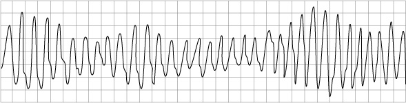

Arrhythmias

Arrhythmias are the main reason for hospitalisation in ACHD patients. Arrhythmias are often a sign of haemodynamic deterioration in CHD patients and they often need urgent Direct Current Cardioversion (DCCV), particularly in the case of patients who have Fontan circulations. Antiarrhythmic drug therapy is often poorly tolerated due to the negative inotropic effects.

Individuals with unexplained syncope (particularly in patients with Tetralogy of Fallot; Transpostion of the Great Arteries and congenitally corrected Transposition of the Great Arteries; Univentricular hearts; and Aortic Stenosis) are at highest risk of sudden cardiac death.


Pulmonary Arterial Hypertention (PAH)

The development of PAH occurs in patients who have untreated systemic to pulmonary shunts (blood flowing through a defect in the heart from the left side to the right side of the heart).

In cases such as Eisenmenger’s Syndrome this shunting process eventually leads to PAH and causes irreversible damage to the lungs which then, in turn, results in a reversal of the shunting. This reversal (blood flowing from the right side of the heart, through a defect and into the left side of the heart) results in the patient becoming cyanosed.

Patients who have PAH in the context of CHD can be offered drug therapies, which need to be managed by a specialist congenital centre. These medications include prostanoids, endothelin receptor antagonists (ERAs) and phosphodiesterase type-5 inhibitors.

Patients with PAH and cyanosis require complex management and the condition will involve multiple organs. Some factors to consider (as per ESC guidelines):

  • High risk of both clotting and bleeding. High risk of cerebral events and abscesses. Should receive prophylactic antibiotics for high risk dental procedures.
  • Important to keep hydrated and avoid acute exposure to heat such as saunas, hot tubs etc. Avoid iron deficiency and anaemia, respiratory infections and agents that can lead to renal impairment.
  • Avoid pregnancy, moderate to severe strenuous exercise and high altitude.
  • Use air filters in IV lines to prevent air embolisms, as high risk of entering system.

Background to congenital cardiac disease

Definition of Congenital Heart Disease:

A cardiac disease or condition that has been present since birth.

Causes

In the majority of cases the cause of a congenital heart condition is unknown. However, there are certain factors which are associated with an increased risk:

  • 17% are associated with other syndromes (Downs Syndrome, DiGeorge and Williams)
  • Certain medications, alcohol or drug abuse
  • Maternal viral infections in the first trimester (such as Rubella)
  • Type 1 and 2 diabetes

Different presentations

Most lesions are detected when the baby is under one years old (60%) and some conditions can be detected before the baby is born, on the mothers 18-20 week foetal scan. However, 10% are not detected until later on in adulthood.

For those adults who are unaware that they have congenital heart disease (CHD), they may present with arrhythmias: syncope or palpitations: chest pain: increasing shortness of breath: heart failure: and/ or hypertension. Some patients present for the first time during pregnancy or their condition is an incidental finding when the patient is having investigations for other reasons.

Prevalence

Historically, babies born with CHD had very poor chances of surviving into adulthood. However, with advances in medical management and paediatric surgery, and with improvements in imaging techniques, such as echocardiograms and MRI, there are now more adults than children with CHD and 96% of babies diagnosed are surviving into adulthood.

Approx 1% of the population, or 1 in every 145 births, will have CHD. Some conditions are more prevalent than others, for example, atrial and ventricular septal defects (ASDs and VSDs) being the most common.

It is estimated that there are approximately 150,000 adults in the UK living with CHD.

Investigations

There are a number of investigations that are used when diagnosing, assessing and managing the treatment of a patient with CHD.

CHD Investigations
ECG Should be routinely carried out at every hospital clinic appointment. 24 hr holter monitors should be used when patients report symptoms of palpitations or there is any suspicion that the patient may have an arrhythmia.
Transthoracic echocardiogram This is the first-line investigation for congenital patients. Ideally, a sonographer should be trained to scan CHD patients.
Transoesophageal echo (TOE) Used during invasive procedures, prior to direct current cardioversion (DCCV) and also in cases where there is a suspicion of infective endocarditis (IE).
Cardiopulmonary exercise test (CPET) Is a tool used to assess base line exercise tolerance and/or detect changes or a deterioration in a patient. It is used to aid decisions around the indication for, and timing of, intervention or surgery. It can also be used a way of measuring the impact an intervention has had on a patient.
Cardiac magnetic resonance imaging (cMRI) Is an increasingly important tool for diagnosis and assessment of CHD patients. It is not suitable, however, for patients who have non MRI compatible pacemakers or ICD’s
Computed tomography (CT scans) Can be used as an alternative for those patients who cannot undergo MRI scans. It is also used as the imaging modality of choice in situations such as assessing patient’s coronary arteries. The high doses of radiation that patients are exposed to will limit the number of scans they are be subjected to.
Right heart catheterisation (RHC) These are carried out in a cath lab and are given to certain patients as part of their assessment. The main purpose of this procedure is to measure the pressures in the four different chambers of the heart, the pulmonary artery and also to assess any shunts that are present across an atrial-septal defect (ASD) or ventricular-septal defect (VSD). The data obtained can assist with planning future medical and/or surgical treatments.
Coronary angiogram This may be undertaken prior to surgery for men over 40 years, post menopausal women and patients with risk factors of coronary artery disease (CAD).

Genetic testing

Gene testing can find:

  • Pathogenic mutations. Other family members can then be offered a gene test.
  • A variant of uncertain clinical significance (VUS). Uncertainty about a VUS result makes a VUS unreliable for diagnosis and this is only used in selected, individual cases.
  • No mutation, therefore, cannot offer a gene test and will not be useful to test other family members.

Genetic testing may:

  • provide confirmation of a familial diagnosis
  • if a pathogenetic mutation is found, can offer predictive testing for close relatives
  • guide discussion around reproductive options
  • guide treatment options
  • help predict the likely severity of the condition

Family tree cascade screening

Cascade (predictive) screening involves testing close relatives of patients diagnosed with inherited cardiac disease, or of a Sudden Cardiac Death victim, by cardiac and genetic assessment. Cascade screening achieves a greater rate of diagnosis than general population screening. Once a diagnosis is confirmed in an individual, testing is extended to first degree and second degree relatives. If relatives test positive, their first and second degree relatives are approached and offered testing, and so on.

Benefits of cascade screening include:

  • exclusion of causative mutation in at risk family members – no further clinical screening required if negative gene test
  • early detection and treatment of inherited heart disease if found to be mutation positive
  • removes continuous uncertainty about whether or not an individual will develop the condition
  • can resume “normal” lifestyle if negative for mutation, for example, intensive sport training
  • result may guide on-going frequency of of screening/cardiac follow up required
  • opportunity to adapt lifestyle if positive for mutation

Genetic counselling

DNA strand

Genetic counselling is a process by which individuals at risk of a disorder that may be hereditary are informed of the consequences of that disorder, the probability of developing and transmitting it and the ways in which this may be prevented or lessened.

The aims of genetic counselling are to help the individual and family to:

  • understand the information about the genetic conditions
  • appreciate the inheritance pattern and risk of recurrence
  • understand the options available
  • make decisions appropriate to their personal and family situation
  • make the best possible adjustment to the disorder or risk

Genetic counselling allows information to be made available to “at risk” relatives, promoting early detection and treatment of inherited heart disease in these individuals. It affords the opportunity to address psychological/social/ethical issues which cannot be managed in a routine cardiology clinic.

Background to inherited cardiac disease

Definition

Inherited Cardiac Conditions (ICC) refers to a group of conditions caused by monogenetic changes (that is, caused by changes in just 1 or very few genes) that affect the heart, its conducting system and muscle structure.

Prevalence

There is, currently, a lack of epidemiological statistics on ICC but estimates put the prevalence at 500:10,000 (see individual conditions described below.)

Inherited Cardiac Conditions:

  • heart muscle conditions which affect the heart muscle and rhythm (cardiomyopathies)
  • heart rhythm conditions which cause problems with the heart rate and rhythm (arrhythmias)

Please enable JavaScript in your browser to see this interactive content.