Primary and secondary hypertension

Hypertension is divided into two forms:

  • Primary (or essential) hypertension (95% of people)
  • Secondary hypertension

Primary Hypertension

  • Primary hypertension has no single known cause but several mechanisms are linked to altered pathways in BP control. These are genetic factors, diet especially increased salt (sodium chloride) intake, obesity, insulin resistance, endothelial dysfunction, chronic excess alcohol, ageing, stress and sedentary lifestyle.
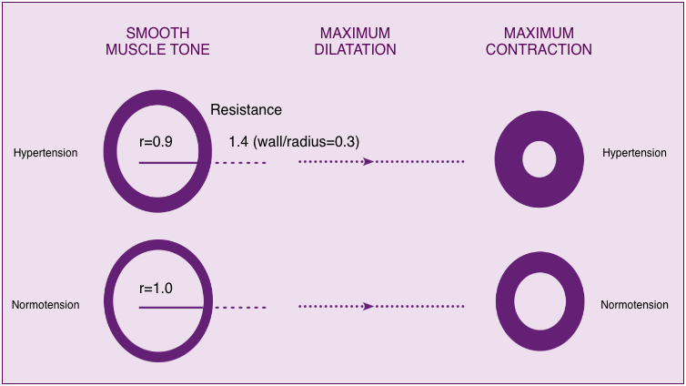
  • The pressure against the blood vessel walls is affected by cardiac output and peripheral resistance. Altered pathways in BP control leads to sustained constriction of the arterioles (microscopic blood vessels in the circulation) resulting in increased peripheral resistance in the blood vessels. As the heart continues to pump normally, the pressure in the whole arterial system rises. This normally has no outward symptoms for the individual, unless very high.

Constricted, stiff arteriole vessel walls lead to increased pressure from blood flow within the arterioles

Arteriole vessel walls

The increased pressure of blood flow against the artery wall leads to damage, resulting in atherosclerotic plaque formation. Signs of prolonged or severe hypertension can be found in target organ damage in the eyes, left ventricle, and kidneys. Presence of target organ damage increases the risk of vascular morbidity and mortality, and the need for treatment to lower blood pressure.

Secondary Hypertension

  • In secondary hypertension BP is raised due to a known underlying cause:
    • Renal disorders (e.g. chronic pyelonephritis, diabetic nephropathy).
    • Vascular disorders (e.g. coarctation of the aorta).
    • Endocrine disorders (e.g. primary hyperaldosteronism).
    • Drugs (e.g. alcohol, cocaine)
    • Miscellaneous causes (e.g. scleroderma, obstructive sleep apnoea).
  • A search for secondary hypertension is only suggested by history, physical examination or routine tests indicate abnormalities.
  • Investigations for secondary hypertension are not cost effective.

What is hypertension?

Hypertension is persistently raised arterial blood pressure (BP) and is a cardiovascular risk factor. 1 in 4 people in the UK have high BP. People with high BP are three times more likely to develop heart disease and stroke and twice as likely to die from these as people with a normal BP (Ref: Department of Health (2001) The Annual Report of the Chief Medical Officer. www.doh.gov.uk). The risk associated with increasing BP is continuous, with each 2 mmHg rise in systolic blood pressure associated with a 7% increased risk of mortality from ischaemic heart disease and a 10% increased risk of mortality from stroke (Ref: NICE 2011 Guideline CG 127 (2011) Hypertension in adults: diagnosis and management).

  • BP is normally distributed in the population.
  • Investigation and management of sustained raised BP is undertaken at levels known to reduce the development or progression of disease, known as thresholds (Ref: NICE 2011 Hypertension The clinical management of primary hypertension in adults Clinical Guideline 127 Methods, evidence, and recommendations http://www.nice.org.uk/guidance/cg127/evidence/full-guideline-248588317).
  • The thresholds for clinic BP readings are:
    • Systolic BP sustained above or equal to 140 mmHg, or
    • Diastolic BP sustained above or equal to 90 mmHg, or
    • Both.
  • The thresholds for Ambulatory BP Measurement (ABPM) or Home BP Measurement (HBPM) are:
    • Systolic BP average above or equal to 135 mmHg, or
    • Diastolic BP average above or equal to 85 mmHg, or
    • Both
  • Diagnosis is based on ABPM (or HBPM) (NICE 2011).

Blood pressure cuff and monitor

  • The greater the level of systolic and/or diastolic BP the greater is the risk of mortality and morbidity associated with
    • Coronary heart disease
    • Stroke
    • Peripheral vascular disease
    • Retinopathy
    • Aortic aneurysm
    • Heart failure
    • End-stage renal disease, or
    • Dementia.
  • Safe, effective, and inexpensive treatment options are available.
  • National guidelines have categorised BP levels and used three stages of hypertension (also known as grades) to aid decision making for diagnosing and offering pharmacological treatment. The same grading is used in young, middle-aged and elderly subjects but not in children or teenagers.
Category Systolic Diastolic
Optimal <120 and <80
Normal 120-129 and/or 80-84
High normal 130-139 and/or 85-89
Stage 1 hypertension 140-159 and/or 90-99
Stage 2 hypertension 160-179 and/or 100-109
Stage 3 hypertension >180 and/or >110
Isolated systolic hypertension >140 and <90

(Table Ref: The European Society of Hypertension (ESH) and European Society of Cardiology (ESC) (2013) ESH/ESC Guidelines for the management of arterial hypertension. J Hypertens 31:1281–1357 DOI:10.1097/01.hjh.0000431740.32696.cc)

Learning outcomes

On completion of this module you will be able to:

  1. Define hypertension and classifications of essential hypertension
  2. Describe the epidemiology of essential hypertension
  3. Explain the pathophysiology of hypertension, including causes and consequences
  4. Outline methods used to confirm a diagnosis of essential hypertension
  5. Consider cardiovascular risk assessment tools used in the assessment of essential hypertension
  6. Appraise the impact of pharmacological treatment and behaviour change approaches
  7. Understand person-centered approaches for the management of individuals living with essential hypertension, focusing upon supporting self-management

Introduction

Introduction - Speech bubble

Module Team

Module Lead

Morven Dunn. Cardiovascular Disease Clinical Development Coordinator, British Heart Foundation

Module Members

James Lambie. Practice Nurse, NHS Lothian
Linda Callan. Coronary Heart Disease Lead Nurse, NHS Lanarkshire
Judith Misson. Health Information Manager, Chest Heart & Stroke Scotland
Susan Kennedy. NHS Education for Scotland, General Practice Nursing

Reviewers

Amanda Manson. Cardiology Specialist Nurse, NHS Orkney
Joanna Toohey. Cardiac Specialist Nurse, NHS Dumfries & Galloway

10. Hypertension

Learning outcomes

  • Define hypertension and classifications of essential hypertension
  • Describe the epidemiology of essential hypertension
  • Explain the pathophysiology of hypertension, including causes and consequences
  • Outline methods used to confirm a diagnosis of essential hypertension
  • Consider cardiovascular risk assessment tools used in the assessment of essential hypertension
  • Appraise the impact of pharmacological treatment and behaviour change approaches
  • Understand person-centered approaches for the management of individuals living with essential hypertension, focusing upon supporting self-management

General Medical Disclaimer: The information, including but not limited to, text, graphics, images, recommendations, opinions and other material contained on or accessed through this website (or in any module or content accessible through this website) (together “Website”), is intended for general informational purposes only. The information should not be considered as professional medical advice, diagnosis, recommendations or treatment.  The information on this Website is provided without any assurance, representations or warranties, express or implied. We do not warrant that the information on this Website is applicable to all healthcare practices, geographical locations, health needs or circumstances. We do not warrant that the information on this module is complete, true, accurate, up-to-date, or non-misleading. You should always seek the guidance of a qualified healthcare professional before making any decisions related to your health or wellbeing. Never disregard or delay seeking medical advice due to something you have read on this Website. The use of or any reliance placed on any information provided on or accessed through this Website is solely at your own risk.  If you have any specific questions or concerns about your health, please consult a qualified healthcare provider or other qualified medical professional. Do not rely on the information on this Website as an alternative to medical advice from your doctor or other qualified professional healthcare professional or healthcare provider.

Module test

Module test certificate icon

This is the module test for ‘HEARTe 9: Cardiac investigations’. It is strongly recommended that you work through the learning materials of the module prior to commencing this test. By going straight to the test you may miss out on valuable learning contained within the module. The answers to all the test questions are contained within the module. This information may have been provided in the ‘Additional Information’ boxes on some of the pages.

There are 10 questions and you must answer all of these correctly to obtain a certificate of completion.

You should allow approximately 10 minutes to complete the test.


Electrophysiological studies (EP Study)

What is an EP study?

An electrophysiology study or EP study is a minimally invasive procedure which aims to map the electrical conduction system of the heart with the aim of identifying the causes of arrhythmias. By identifying the location of the patient’s arrhythmia, a treatment plan can be formulated.

An EP Study is often an elective procedure carried out in a specialist Cardiac Catheter Laboratory in a hospital. It usually requires the patient to stay for one night in hospital. It is carried out by a team including a cardiologist, cardiac physiologists, radiographers and nurses.

The Cardiac Catheter Lab layout will differ between hospitals. The X Ray machine usually arches over the patient and can provide images of the heart and the catheter placements during the procedure.

The EP Study:

  • may take around 2-3 hours and sometimes longer
  • The patient will be taken to the Cardiac Catheter Lab and made as comfortable as possible on an X Ray table
  • At the start of the procedure the patient will often be offered a sedative through an IV cannula to help them relax
  • Once comfortable and medication administered, the team will then start connecting the patient to monitoring equipment

The cardiologist and cardiac physiologists may already have knowledge of the patient’s arrhythmia which will dictate the equipment used during the procedure. Standard monitoring equipment will be applied such as BP cuff, SpO2 probe and 3 lead ECG. If appropriate, defibrillation pads may be connected at the start of the procedure if the patient’s arrhythmia is know to be unstable/life threatening.

A local anaesthetic will be injected at the catheter entry sites. The entry sites are usually at the right groin and right wrist to gain access to the right femoral vein and the right radial artery or a vein in the right forearm.

The cardiologist will start the procedure and ‘map’ a picture of the heart and electrical conduction system using the catheters. The X Ray machine allows the cardiologist to locate the chambers of the heart

Below are two examples of diagnostic catheters. As you can see they are very flexible and have electrodes along the outside and on the tip:

EPS catheters

There may be 3 to 5 catheters used during a procedure. The catheters send data to the cardiac physiologists and it is interpreted in graphics an ECG waveforms for the cardiologist. The electrodes on the catheters can detect the electrical activity in the heart. When an arrhythmia is detected the cardiologist can see where it is present and can make a treatment plan from there.

During the procedure, the cardiologist may identify an arrhythmia which can be treated with ablation therapy, where radiofrequency energy is used to destroy or freeze the particular area of heart tissue that is the source of the arrhythmia. Arrhythmias which can be treated by ablation are:

  • Supra-ventricular tachycardia
  • Wolff Parkinson White
  • Atrial Flutter
  • Atrial Fibrillation
  • Atrial Tachycardia
  • Ventricular Tachycardia

It is essential that emergency resuscitation equipment is readily available during this procedure due to the arrhythmias which may be precipitated by the test.

Other treatment options following an Electrophysiology Study include medication therapy, a pacemaker, an implantable cardioverter defibrillator (ICD) or cardiac surgery.

See Additional Information for a BHF patient video.

Other monitoring

Other monitoring

Having covered cardiac monitoring and 12-lead ECGs, we will now consider other types of cardiac monitoring/investigations which may be performed. This section will include a variety of ambulatory electrocardiography (AECG) tests and the electrophysiological (EPS) study.

For a person presenting with palpitations, dizziness, near-syncope (feeling that they are going to pass out) or syncope (episode of loss of consciousness), we need to determine which type of ECG test will most likely help provide a diagnosis.

Which test?

To determine which type of ECG test is appropriate, it is essential:

  • To seek a clear description of the symptoms:
    • how long have symptoms been occurring
    • the number of times symptoms have occurred in the past year
    • the amount of time between events
    • any injuries sustained during events
    • any eye-witness account
    • any warning signs
  • To consider the clinical context:
    • does the patient have underlying coronary artery disease
    • does the patient have underlying structural heart disease

Types of ECG tests:

  • Continuous ambulatory ECG monitoring: Holter monitoring
  • Intermittent ambulatory electrocardiography (AECG)
  • Implantable Loop Recorder (ILR)
  • Resting 12-lead ECG
  • Exercise tolerance test

Please enable JavaScript in your browser to see this interactive content.

See Additional Information for BHF patient videos for ILRs and Holter monitoring in use.

George

George is a 59-year old diabetic man who has noticed he is becoming more breathless. He is too worried to go to his doctor in case there is something serious.

After several months he cannot walk far without being out of breath and his ankles are swollen.

Please enable JavaScript in your browser to see this interactive content.