Tilt table testing

Tilt testing is used to investigate episodes of unexplained syncope when a cardiac arrhythmia is unlikely. Syncope comes from the Greek ‘to interrupt’ and is a common medical problem.

Syncope occurs when there is transient global cerebral hypoperfusion ( not enough blood flow to the brain). This can be either due to low blood pressure or a heart problem.

When a patient presents with syncope the evaluation should include:

  • History – including eye witness accounts
  • Examination of the heart
  • 12 lead resting ECG
  • Lying and standing BP

There are 4 main types of syncope:

  1. Neurally mediated is the most common cause and refers to a fainting reflex (vasovagal) which is triggered in some people by certain situations e.g. sight of blood.
  2. Orthostatic (postural) hypotension (OH). In older people low blood pressure and orthostatic hypotension is much more common than neurally mediated syncope. In OH the blood pressure falls immediately on standing due to impaired autonomic reflexes. It is common in older people and can be exacerbated by drugs or other diseases e.g diabetes. There is another common pattern in older people called ‘elderly dysautonomic pattern’ where there is a slow fall in BP after standing which goes undetected on normal lying and standing BPs.
  3. Cardiac arrythmias account for around 20% of syncope.
  4. Structural causes for example aortic stenosis or hypertrophic obstructive cardiomyopathy (HOCM).

Tilt testing aims to reproduce what happens when a patient has neurally mediated or OH syncope at home.


How a Tilt Table Test is performed

Please enable JavaScript in your browser to see this interactive content.

The test is performed as an out patient in a hospital clinic and it is usually supervised by two people, a cardiac physiologist/doctor/specialist nurse trained in life support and an assistant. Resuscitation equipment is immediately available and a doctor nearby should any problems arise, although the test itself is extremely safe.

Rarely patients may develop arrhythmias. For example young people with neurally mediated syncope( especially with situational syncope) may get transient asystole, but this is what would be happening to them at home when they faint.

The patient may be asked to fast before test, to empty their bladder, undress to the waist (the patient will be given a gown to wear) and remove any support stockings before the test.

Ideally the test should be done in a quiet room with dimmed lighting. The patient then lies flat on a special table that can be tilted at various degrees to an upright position. The patient has their feet against a foot board and is strapped on to prevent them slipping off.

The test lasts for approximately 40mins. Patients are continuously monitored with 12lead ECG, continuous non invasive beat to beat blood pressure monitoring and intermittent blood pressure using Dynamap or similar.

Blood pressure and heart rate are recorded every 2 minutes. The patient is tilted to 60-70degrees for 20mins, if no change in HR or BP, tilted for another 20mins (Dependant on local protocol GTN spray may be given to stimulate a reaction). If the test end points have not been reached then the test is concluded.

See “Additional Information” for a patient video.


Termination of test

The test will be stopped and the patient laid flat immediately if:

  • Systolic blood pressure falls below 80mmHg or is falling rapidly
  • Heart rate falls below 50/min, or is falling rapidly
  • Heart rate rises above 170/min
  • Acute arrhythmia occurs
  • Hyperventilation occurs
  • The patient is distressed or uncomfortable
  • The end of protocol has been reached

Response to tilt testing

A positive tilt test is when the patient’s syncopal or pre-syncopal symptoms are reproduced and accompanied by :

  1. Bradycardia
  2. Hypotension or
  3. Both

There are several different positive responses to a tilt test:

  • Vasodepressor – BP falls but the heart rate does not.
  • Cardio-inhibitary A ( without asystole) –HR falls to less than 40/min for more than 10seconds but asystole is less than 3 seconds.
  • Cardio-inhibitary B ( with asystole) – there is asystole for more than 3 seconds.
  • Mixed – a mixture of vasodepressor and cardio-inhibitary response A.
  • Excessive HR rise – if the heart rate rises at onset of upright position to > 130/min and throughout test before syncope occurs, this is known as POTS- postural orthostatic tachycardia syndrome.
  • Chronotropic incompetence – there is no HR rise during the test i.e < 10% from baseline.

Cardiac magnetic resonance imaging (Cardiac MRI)

MRI scanner

Cardiac Magnetic Resonance Imaging (Cardiac MRI) is a non-invasive test which allows the structure and function of the heart to be studied in detail, without the need for radiation.


What the test involves

Cardiac MRI is performed in a MRI scanner, often situated in the radiology or cardiology department. The MRI scanner is essentially a big, high strength magnet which uses different coils to help generate images. On arriving at the scanning department, a radiographer will meet the patient and go through a safety questionnaire to ensure that it is safe for the patient to enter the MRI scan room. A Venflon (vascular cannula) will be inserted into an arm vein. The patient will be asked to change into a hospital gown and lie down on their back on the scanning table. ECG leads will be attached to the chest to help monitor the different parts of the cardiac cycle. A cardiac coil will also be placed on the chest, to help generate the images. Once the ECG leads and cardiac coil are in place, the scanning table will be moved into the MRI scanner. The scanner makes a series of loud noises, and earphones are usually put on the patient to mask the noise. Music can be played through these to mask the noise and help relaxation. The patient can be seen and heard at all times by the staff performing the scan, by a live video link and by a microphone.

The radiographer will perform a number of scan sequences. Depending on the question being asked by the referring doctor, this series of scans may take up to one hour. The get the best image possible, the patient must lie very still. He/she will also be given breathing instructions prior to each sequence (breathe in, breathe out, hold your breath). Some breath holds may be up to 15 seconds.

During some cardiac MRI scans, a contrast agent called Gadolinium will be given intravenously. Using Gadolinium allows an assessment of cardiac perfusion (blood supply) to be made as well as identification of scarring or fibrosis in the heart. This is given as a painless injection through a Venflon and it can sometimes feel cold as it travels up the arm. If the radiographer knows that Gadolinium will be required during the cardiac MRI, the patient will have had a blood test prior to the scan to ensure kidney function is satisfactory. Most but not all patients require Gandolinium.

Once the scan has been completed, the patient’s Venflon will be removed prior to the patient going home (or back to the ward). The result of the scan will not be available immediately, as it required to be analysed in some detail by either a cardiologist or radiologist (or both). The result will go back to the requesting clinician.


Which patients will the test be used on and why?

Cardiac MRI may be used for many different reasons. It is used to study both the anatomy (structure) and the function of the heart. Some common reasons for asking for a cardiac MRI are:

  • To get an accurate assessment of ventricular function (how well the heart chambers are contracting)
  • To look at valve structure and function
  • To assess whether there is any problem with the blood supply to the heart
  • To assess the major blood vessels (eg aorta)
  • To assess the pericardium (the sac around the heart)
  • To measure the thickness of the heart muscle
  • To assess the degree of scarring after a heart attack, and whether the heart muscle might recover if blood supply is restored
  • To assess congenital heart disorders
  • To assess tumours in or around the heart

How is it used to diagnose cardiac conditions?

Once the scan has been performed, a cardiologist or radiologist (or both) will review the scan and produce a report for the requesting doctor. Patients will not normally receive the result at the same time of the scan.

CT Coronary Angiogram

Definition of Test

A computerised tomography coronary angiogram (CTCA) is a non-invasive method of imaging the coronary arteries. Unlike invasive coronary angiography, it does not require the use of catheters although does still involve X-rays. The test requires a 64 detector (or higher) CT scanner and involves an intravenous injection of an iodine-based dye to generate high resolution 3D images of the coronary arteries. The higher the number of detectors on the CT scanner, the better the image. The scanner takes multiple images of the heart from various different angles. The images obtained allow detection of fatty plaques or calcium deposits in the coronary arteries, which can result in coronary artery narrowing or blockages.


What the Test Involves

To obtain a clear picture of the coronary arteries in this test, it is important that the heart is not beating too quickly. Ideally, the heart rate should be 60bpm or less. Many patients will have been asked to take medication on the morning of the test to slow the heart down (eg beta-blockers, calcium-channel antagonists or ivabradine). On arriving for the test, the patient will be asked to change into a gown and an intravenous cannula inserted. Heart rate and blood pressure will be checked. If the heart rate is too fast, further medication will either be given by mouth or by injection into an arm vein, to slow the heart further. Once the heart rate is slow enough, the scan will be performed.

The patient will be asked to lie on a bed whilst ECG electrodes are placed on the chest. This allows the heart beat to be monitored during the test, and also allows us to ensure that we scan the heart during the same part of the cardiac cycle each time. The bed will then move into the scanner. See “Additional Information” for a patient video.

Most centres will start with an unenhanced scan (ie one that does not require the use of contrast to enhance the images). This is a very quick scan which detects the presence of calcium deposits in the coronary arteries (CT-calcium scoring). If there is a large amount of calcium in the coronary arteries, it can degrade the quality of the scan and make it non-diagnostic and for this reason patients with a very high calcium score may have the procedure terminated at this point. Calcium scoring is measured by the Agatston Score. A value of >400 is usually taken as ‘severe’ calcification and may result in the scan being terminated in favour of an alternative method of assessing the coronaries (eg an invasive coronary angiogram).

For those patients with a lower calcium score, an intravenous injection of an iodine-based dye is then given to help further visualise the coronary arteries. It is usually necessary to breath-hold for a few seconds during this part of the scan, to minimise movement of the heart and lungs. X-rays pass through the body and are picked up by detectors in the CT scanner, which then generates an image of the coronary arteries.

The scan itself usually takes 15 minutes or less, although there may also be some waiting about afterwards for monitoring if extra heart-slowing medication has been given.


Which patients might a CTCA be used on and why?

CT coronary angiography is best used as a ‘rule out’ test for coronary artery disease. The sensitivity for CT coronary angiography is high at 89% and the specificity even higher at 96%. The high level of specificity is supported by a high “negative predictive value” which indicates what a reliable test CT angiography is for ruling out coronary artery disease if the test is negative i.e. normal.


Common reasons for requesting a CT coronary angiogram include:

  • For the evaluation of coronary artery disease in stable, low risk chest pain patients (as recommended by NICE Clinical Guideline 95) seen either in rapid access chest pain clinics, cardiology clinic or as inpatients
  • To visualise coronary arteries in patients with heart failure of unknown cause.
  • To evaluate coronary artery bypass grafts.
  • To investigate patients who may have anomalous (abnormal) origins of coronary arteries
  • To assess coronary arteries in patients with endocarditis and aortic root abscess, prior to valve surgery. In such patients, manipulation of an invasive catheter within the aortic root might be undesirable and a CTCA safer.

How is CTCA used to diagnose a cardiac condition?

Once the images have been acquired, they can be viewed on a computer by either a radiologist, a cardiologist or both. Post-processing allows 3D reconstruction of the coronary arteries to be performed. A report will then be issued to the requesting doctor. Patients are not usually informed of the result on the day of the test.

Benefits of CT-A over invasive coronary angiography:

  • No requirement for invasive vascular access
  • No risk of stroke/MI/vascular damage

Coronary angiography

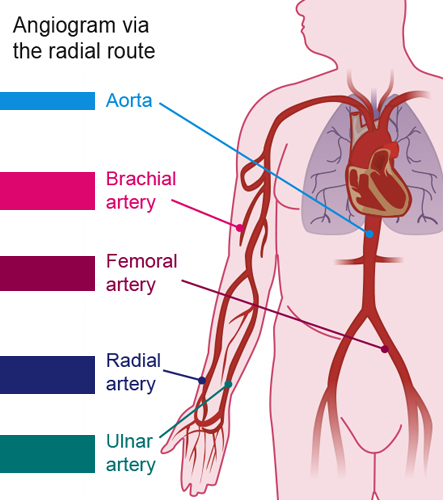
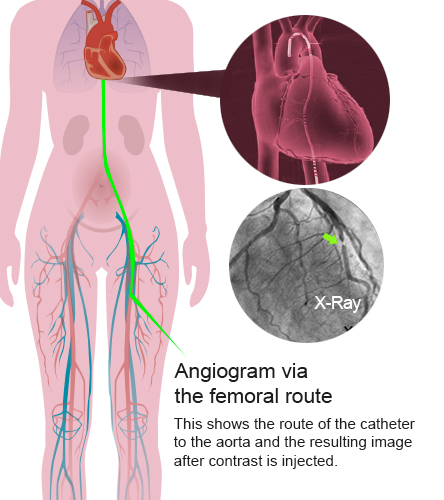
Angiogram via radial routeAngiogram via femoral route


Coronary angiography is a direct method of imaging the coronary arteries. This invasive procedure involves introducing fine cardiac catheters (approximately 2mm in diameter) inserted over guidewires (Seldinger technique) into the radial (or occasionally femoral) artery and advanced to the aorta. The catheters are designed to select the left and right coronary arteries. X-ray contrast medium (usually Iodine based) is then injected and radiographic images taken of the coronary arteries to visualise areas of coronary stenosis or occlusion.

This can determine the need for and best mode of revascularisation in patients with coronary artery disease. See Acute Coronary Syndrome: Case 1 Hamish for an animation of a coronary angiogram performed via the right radial approach and a Video of a coronary angiogram showing 3 vessel disease.


Risk of MI, Stroke or Death

  • Risk is estimated at < 1 in 1000 (Ref-British Heart Foundation 2014) However the level of risk for the individual depends on their overall health, age and heart condition.
  • The Clinician will not recommend the test unless it is believed that the benefits outweigh the risk, however he/she must discuss this fully and openly with the patient.
  • The patient must give written informed consent prior to the procedure.

Patient information before the procedure:

  • Patients should be advised they may feel a bit of pushing and pulling as the catheter is manoeuvred into place and a warm flush feeling as the dye is injected
  • They will be given local anaesthetic at the entry site (wrist or groin)
  • They can be given some light sedation if anxious about the procedure

Safety Issues & Complications

Coronary angiography is relatively safe however it is an invasive procedure and there are some risks of complications:

  • Insertion of the guide wire can cause arterial trauma
  • Air bubbles in the cardiac catheter can cause air embolus
  • Haematoma can occur at the arterial access site (more common when the femoral artery is used)
  • Embolisation of thrombus or atheroma is possible
  • Arrhythmias (atrial or ventricular) are possible during the procedure
  • A vaso-vagal response may occur during the sheath removal from the femoal atery
  • Coronary occlusion
  • Dye sensitivity can cause nausea, hypotension or urticarial N.B It is important to check first if patients have any allergies – to previous dye, iodine or shellfish (contain iodine)
  • The contrast can be nephrotoxic therefore there is a risk of renal failure – increased risk if the patient already had renal impairment
  • Having a coronary angiogram involves exposure to some radiation, which is more than a CXR but less than an MPS or CT coronary angiogram.

Please enable JavaScript in your browser to see this interactive content.

Myocardial Perfusion Scintigraphy (MPS)

Definition of the test

A myocardial perfusion scan (also known as myocardial perfusion scintigraphy) is a non-invasive nuclear medicine imaging test which uses a small amount of a radioactive tracer to investigate the blood supply to the heart. It is a functional test which can detect any difference in blood flow to the heart muscle at rest and during stress (which may be exercise stress or pharmacological stress). It can also be used to assess the extent of damage to the heart after a heart attack. It does not look at the coronary arteries themselves; rather it looks at the blood supply to the heart muscle, from which inferences can be made about the arteries.

The requirements for a MPS include a radioactive tracer, a stressor and a gamma camera. The radioactive tracer (which may be technetium, thallium or others) emits radioactivity in the form of gamma rays. Once it is injected into a vein, it travels in the bloodstream and is taken up by heart muscle. Areas of the heart that have a good blood supply will take up a large amount of tracer, whereas areas of the heart with a poor blood supply will take up a smaller amount of tracer. Therefore, areas of heart muscle with a good supply will emit more gamma rays than areas with a poor blood supply.

Radiation emitted by the tracer is detected by a gamma camera, and converted into an image on a computer.

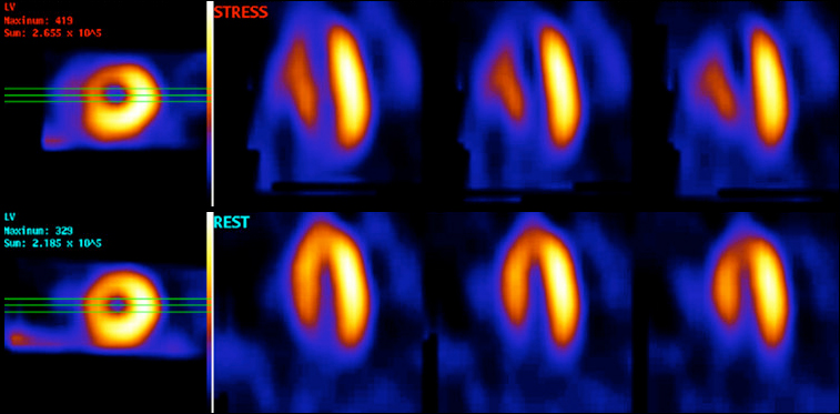
MPS stress and rest
The top row shows stress images, bottom row shows rest images. This patient has a stress-induced perfusion defect in the apex and septum (probably LAD disease)

What it involves

There are a number of different protocols for performing a MPS, which vary from hospital to hospital. Many centres utilise a two part scan, whereas some perform the whole scan on one visit. The test will involve a ‘stress’ part and a ‘rest’ part, to allow comparison of blood flow during stress and rest.

If the planned test involves pharmacological stress, patients will have been asked to withhold/avoid certain medications or drinks containing caffeine for a day or two prior to the appointment. If this is required, the patient appointment letter will usually contain this information.

On arrival at the nuclear medicine department, the patient will be prepared and consented as for any invasive procedure. See ‘Additional Information’ for a patient video on this test.

During the stress part of the test, the heart is made to work harder either with gentle exercise on a treadmill or bicycle, or alternatively with infusion of a drug. Some common drugs that might be used are those which make the heart beat stronger and faster, such as dobutamine, or those which vasodilate (open up) the arteries and increase blood flow to the heart, eg adenosine or dipyridamole. Adenosine and dipyridamole are generally not used in patients with reversible airways disease such as asthma, as they can cause bronchoconstriction and breathlessness. Once the stressor has been given, the radioactive tracer will be injected and the patient will be asked to lie in the scanner to allow the gamma camera to detect the emitted gamma rays. Patients are usually required to place their arms above their head whilst the scan is being performed, and lie as still as possible. The scan can take up to 30 minutes.

The stress scan images will generally be reviewed by a nuclear medicine consultant, radiologist or cardiologist and a decision made as to whether the patient requires to return for a ‘rest’ study. This will depend upon the reason for the test and whether there are any abnormalities on the stress study. If the patient requires to return for a rest study, this will involve an injection of the radioactive tracer followed by a scan – this part is usually much quicker, as there is no need for exercise or pharmacological stress.


Which patients might a MPS be used on and why?

MPS is used to identify whether there is a problem with the blood flow to the heart muscle, or to assess how much damage has been done to the heart after a myocardial infarction. It is therefore of use in patients:

  • With chest pain, to help diagnose or rule out angina
  • With an abnormal coronary angiogram, to help identify which narrowed artery is likely to be the culprit for angina (and therefore which artery should be treated).
  • With impaired left ventricular function on echo – to assess whether the areas of the heart which are not contracting well are still viable (and worthy of restoring the blood flow to) or not.

How is MPS used to diagnose a cardiac condition?

When assessing blood flow to the heart in a myocardial perfusion scan, we look at whether there is a difference in regional myocardial blood flow at rest and following stress. There are three main possibilities:

  1. If the blood supply at rest is normal, but the blood supply following stress is reduced, this indicates that there is reversible myocardial ischaemia which is likely to be due to a narrowing in a coronary artery. This is called a reversible perfusion defect. By comparing the tracer uptake in the various different areas of the heart (the coronary artery territories) we can work out which artery or arteries are likely to have a narrowing. Such patients might go on to have an invasive coronary angiogram to identify the narrowings and potentially treat them.
  2. If blood supply at rest is the same as blood supply following stress, it is unlikely that there is significant obstructive coronary artery disease. This is a normal myocardial perfusion scan. For patients with chest pain, a normal myocardial perfusion scan is associated with a rate of MI or cardiac death of less than 1% per year. It is therefore a good ‘rule out’ test for significant obstructive coronary disease.
  3. If blood supply at rest is abnormal but is similar to the blood supply following stress, this suggests that there has been previous myocardial infarction in the area of reduced blood supply. This is called a fixed perfusion defect. Such fixed defects can represent either viable or non-viable myocardium – the former might recover function with revascularisation whereas the latter will not.

The stress and rest images will be reviewed by a nuclear medicine consultant/cardiologist/radiologist and a report generated for the referring doctor. Patients will not normally receive the result on the day of the scan.

Benefits Risks
  • Non-invasive way of assessing likelihood of having significant coronary artery disease
  • Test tolerated very well in most cases
  • Involves small amount of radiation
  • Some patients find the pharmacological stressor unpleasant

Stress echo

A stress echocardiogram is usually done on low cardiac risk patients with unexplained chest pain or dyspnoea, which has been triggered by physical activity.

Non pharmacological stress echocardiogram

This is an echocardiogram carried out during, or just after, a period of exercise on a treadmill or exercise bike, usually to assess mitral or aortic valve function, or assess left ventricular outflow tract velocities if hypertrophic cardiomyopathy is suspected.

Pharmacological Stress Echo

For those patients who may have had unequivocal result from a non pharmalogical stress echo or may be physically be unable to do one, a pharmalogical stress echo may be carried out. A pharmalogical echo may also be used in patients with known coronary artery disease and left ventricular dysfunction, to help identify whether intervention to the arteries might help heart function (viability study). Similarly, in patients who have aortic stenosis and left ventricular impairment, a low-dose dobutamine study can help us decide how severe the aortic stenosis actually is.

The test is used to visualise the effect of increasing heart rate on the contraction of the left ventricle and determine whether there is reduced blood flow to the myocardium, producing ischaemic changes on the scan and ECG.

A Dobutamine infusion (or occasionally Dipyridamole depending on information required) is used to artificially increase the heart rate. An echo is then performed at pre determined stages. This is usually pre Dobutamine; at low dose; peak heart rate; and in recovery stage.

The test involves the patient being cannulated and attached to an injection pump containing a saline/ Dobutamine mix. 12 lead ECG and BP are continuously monitored throughout the test.

  • Benefits:
    • Instant results
  • Risks:
    • Hypotension
    • Arrythmia: AF, VT
    • Chest pain: causing ischaemia or M.I

Dobutamine Stress Echo (DSE) is generally done within a specialised department, with appropriate resuscitation equipment and medications close at hand. A specialist nurse, cardiologist and echo physiologist are present throughout.

The infusion is run until the desired heart rate is reached. This is the same as an Exercise Tolerence Test (ETT). It is calculated as 220 beats per min (minus) – the patients age, x 0.85%. Atropine may be added if the heart rate response is inadequate.

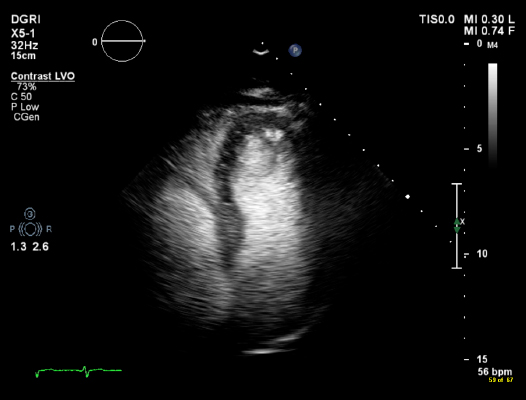
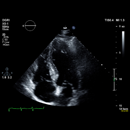
A special contrast agent containing microbubbles of sulphur hexafluoride is injected to enhance the echo images by defining and outlining the LV cavity and myocardium. Sonovue contrast is the most commonly used contrast agent in the UK. This gas has a very short life and is absorbed by the lungs very quickly, and has no effect on the patient.

The full test lasts approximately 40 minutes and the results accurately analysed.
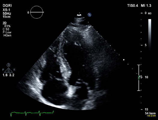
Stress echo 1

Same image with contrast medium

Stress echo 2

Transoesophageal echocardiogram

A Transoesophageal Echocardiogram (TOE) is an invasive procedure, where a transducer similar to an endoscope is passed through the mouth, down the throat, into the oesophagus and stomach to allow very clear and unobstructed imaging of the heart structures. It is done mainly to look closely at the valve structure and function, and assess the inter-atrial septum. The patient can have this procedure as an in-patient or electively as an out patient, however, the process is the same. The results are usually instant and often the patient is told at the time of procedure.

The procedure is normally quite straightforward, taking about 20 minutes. There are some risks involved, such as tearing of the oesophagus, damage to teeth/ mouth or breathing difficulties. The patient would be prepared and consented as for an invasive procedure. Cardiac monitoring and vital signs are continuously recorded during the procedure. The patient may be offered sedation as the procedure may be uncomfortable.

The patient will usually have had a standard transthoracic echo previously and the TOE is carried out to help assess and diagnose any problems that the transthoracic echo alone did not answer.

Indications Contraindications
  • Thorough native and prosthetic valve assessment prior to or during cardiac surgery.
  • Detection of intracardiac thrombus in left atrial appendage prior to cardioversion for atrial flutter/ fibrillation.
  • Detection and visualisation of intra-cardiac shunts.
  • Cardiac source of embolus.
  • Assessment of thoracic aorta/root
  • Good visualisation of aortic root where abcess may be suspected.
  • Assessment of intra-cardiac masses.
  • Oesophageal varices
  • Oesophageal obstruction or stricture
  • Recent gastric or oesophageal bleeding
  • Severe respiratory depression
  • Severe coagulopathy
  • Cervical spine injury or anomaly

How is it done?

The patient will be asked to fast for 6-12 hours before the procedure. Some centres will ask the patient to stop taking anti-coagulants such as Warfarin before the TOE.

See Additional Information, below, for a patient video on how the test is performed.

Why do an echocardiogram?

An echocardiogram can help diagnose and monitor many heart conditions by checking the structure and function of the heart and blood flow through the heart valves and surrounding blood vessels. By using high frequency sound waves ( Doppler) it is possible to assess how the blood flows through them and to assess the pumping action of the heart.

An echo cannot show the coronary arteries that supply the heart, as they are too small to be imaged by ultrasound.

Please enable JavaScript in your browser to see this interactive content.

What is an echocardiogram?

An echocardiogram, or “echo”, is a type of ultrasound scan used to look at the heart structure, primarily the valves and ventricular function. The most common form of echo is transthoracic (TTE). Depending on the type of information required, a more invasive procedure namely transoesophageal echo (TOE) may be performed.

A transducer emits high-frequency sound waves that create returning echoes when they bounce off heart structures and blood cells. These returning echoes are picked up by the electronic crystals in the probe and turned into a moving image that is displayed on a monitor while the scan is carried out.

Please enable JavaScript in your browser to see this interactive content.

The test will normally be carried out at a hospital or clinic, by a cardiologist or a cardiac physiologist. For basic details on an ECHO of a normal heart see Common Cardiac Investigations: Echocardiogram (ECHO).

How a TransThoracic Echocardiogram (TTE) is carried out

For a TTE, ideally, the patient will be asked to lie on their left side. This brings the heart forward in the chest cavity, enabling better imaging between the ribs and around the lungs. An ECG is attached to help allow the operator to assess the rhythm and as an aid in making the correct measurements. Ultrasound gel is used as a transmitter and the probe is placed in several different positions around the chest to enable the operator to see as much as possible.

The procedure is generally pain free but can be a little uncomfortable.

The whole procedure will normally take between 15 and 30 minutes depending on the information required, the complexity of the study required and the ease with which the images are seen.

Benefits of echo Disadvantages
Instant analysis of images can aid clinician on medical or surgical decision making

There are no known risks with this procedure

  • Time consuming
  • Requires experienced operator
  • Quality of images dependant upon good echo subject e.g poor imaging in obese people/ COPD/ chest trauma or radiotherapy and further more complex imaging may be required such as transoesophageal echo or cardiac MRI.

The following case study 04: Acute Coronary Syndrome: Case 2: Joan shows the use of echocardiography in clinical practice.

See Additional Information,below, for a British Heart Foundation video of a patient undergoing a echocardiograph.

Imaging

Imaging featured

Introduction

In this section, which expands on Common Cardiac Investigations, we are going to cover the various types and methods of cardiac imaging to look at the structure and function of the heart.

Some of these tests are highly specialised and are only done in tertiary centres, while others can be done readily in district general hospitals or clinic settings. This will include non-invasive imaging, such as echocardiography, and invasive imaging, including coronary angiography, computerised tomography, coronary angiography (CTCA) and myocardial perfusion imaging (MPI).

The clinician will decide which imaging modality is most appropriate for the patient, depending on the information required.

If the patient is experiencing breathlessness or chest pain, commonly the first test to be asked for is a Transthoracic echo (TTE). This is used to look at left ventricular function and rule out any underlying structural abnormalities. For various reasons, possibly due to their chest shape, previous chest surgery, previous radiotherapy, COPD or high BMI some patients are not deemed echogenic and transthoracic imaging is poor. In these cases a transoesophageal echo (TOE) may be used.

For visualising LV function, sometimes the addition of an echo contrast medium is enough to give good images. This can be a factor in making a clinical decision regarding appropriateness of which imaging to use.

Depending on the results of the TTE the patient may be referred for a more specialised form of imaging. These can all be used in the assessment of LV function:

  • Dobutamine stress echo
  • MPI
  • Magnetic resonance imaging (MRI)

Other forms of imaging can be used to look directly at the coronary arteries:

  • CTCA
  • Angiography