The GP suspects that Andrew’s presenting symptoms are suggestive of a cardiac condition and urgently refers him to the nearest cardiology clinic. Andrew attends the clinic, accompanied by his father, where he undergoes the following tests:
Please enable JavaScript in your browser to see this interactive content.
INTERACTIVE IS FULL OF LINKS
Based on the results of his investigations, Andrew is given a diagnosis of hypertrophic cardiomyopathy.
What is hypertrophic cardiomyopathy?
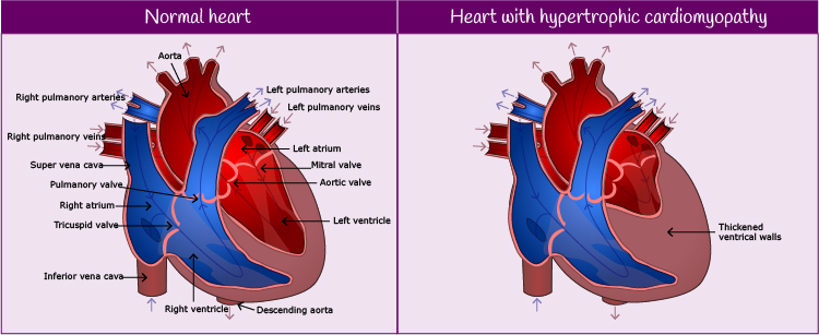
Hypertrophic cardiomyopathy occurs when the cells in heart muscle enlarge and cause the walls of the ventricle (normally the left ventricle) to thicken. It can be diagnosed at any age and is more common in men than in women. For more information on hypertrophic cardiomyopathy, please see Additional Information, below.
Andrew undergoes the following tests:
- An ECG demonstrated that Andrew was in sinus rhythm but has Left Ventricular Hypertrophy. See Cardiac Investigations module: Electrocardiography for more information.
Video: British Heart Foundation – Your guide to ECG (electrocardiogram), heart disease test - A Transthoracic Echocardiogram showed evidence of hypertrophic cardiomyopathy. See Cardiac Investigations module: What is an echocardiogram? for more information.
British Heart Foundation – Your guide to an Echo (echocardiogram), heart disease test - A Holter ECG was used to look for underlying arrhythmias but no abnormal changes were detected. See Cardiac Investigations module: Other monitoring for more information.
- A Loop recorder was then implanted, which showed episodes of non-sustained ventricular tachycardia. See Cardiac Investigations module: Other monitoring for more information.
- Exercise Tolerance Test: may show exercise induced VT. See Stable Coronary Artery Disease: Case 2 Mary for a video.
- An MRI may be performed to detect thickening of the ventricular wall in areas that are difficult to assess by echocardiogram. See Cardiac Investigations module: Imaging: MRI for more information.
Video: British Heart Foundation – Your guide to a cardiac MRI, heart disease test - Genetic testing for sarcomere gene mutations (known to be present in hypertrophic cardiomyopathy) may be undertaken. See Inherited & Congenital Cardiac Disease: Introduction: Genetic testing for more information.
Pulse point
There are a number of different formulae for diagnosing LVH via ECG. One of these is the Sokolow-Lyon formula:
- S in V1 or V2 R in V5 or V6 (whichever it is the larger) ≥ 35mm (≥ 7 large squares)
- R in aVL ≥ 11mm