The paramedics arrive and perform a 12-lead Electrocardiogram (ECG). This shows left bundle branch block (LBBB). The ECG is telemetred to the Royal Heart Infirmary who advise transfer to the cath lab for immediate primary angioplasty. The paramedics are told that Naveed has received aspirin 300mg, so give him clopidogrel 600mg (both orally) and heparin 5000 units (iv) after establishing intravenous access. Oxygen is administered as Naveed’s saturations are low.
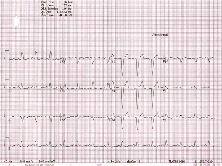
Left bundle branch block is caused where there is delayed depolarisation of the left ventricle. On the ECG this is seen as a lengthening of the QRS complexes. It can be a sign of an acute ST elevation MI.
Pulse point
There are other possible causes for LBBB but the finding of new LBBB in the context of cardiac sounding chest pain suggests that the patient is suffering from an ST Segment Elevation Myocardial Infarction(STEMI). For many patients it will not be known whether LBBB is old or new and a clinical judgement has to be made as to whether the patient is having a heart attack or not.It is good practice to give patients with known LBBB a copy of their ECG or a card to help decision making in emergency situations.
You may want to review Module 1 ECG information or access the following resource Patient.co.uk – ECG A Methodical Approach
Page last reviewed: 12 Jun 2020