Cardiac Magnetic Resonance Imaging (Cardiac MRI) is a non-invasive test which allows the structure and function of the heart to be studied in detail, without the need for radiation.
What the test involves
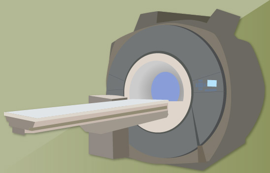
Cardiac MRI is performed in a MRI scanner, often situated in the radiology or cardiology department. The MRI scanner is essentially a big, high strength magnet which uses different coils to help generate images. On arriving at the scanning department, a radiographer will meet the patient and go through a safety questionnaire to ensure that it is safe for the patient to enter the MRI scan room. A Venflon (vascular cannula) will be inserted into an arm vein. The patient will be asked to change into a hospital gown and lie down on their back on the scanning table. ECG leads will be attached to the chest to help monitor the different parts of the cardiac cycle. A cardiac coil will also be placed on the chest, to help generate the images. Once the ECG leads and cardiac coil are in place, the scanning table will be moved into the MRI scanner. The scanner makes a series of loud noises, and earphones are usually put on the patient to mask the noise. Music can be played through these to mask the noise and help relaxation. The patient can be seen and heard at all times by the staff performing the scan, by a live video link and by a microphone.
The radiographer will perform a number of scan sequences. Depending on the question being asked by the referring doctor, this series of scans may take up to one hour. The get the best image possible, the patient must lie very still. He/she will also be given breathing instructions prior to each sequence (breathe in, breathe out, hold your breath). Some breath holds may be up to 15 seconds.
During some cardiac MRI scans, a contrast agent called Gadolinium will be given intravenously. Using Gadolinium allows an assessment of cardiac perfusion (blood supply) to be made as well as identification of scarring or fibrosis in the heart. This is given as a painless injection through a Venflon and it can sometimes feel cold as it travels up the arm. If the radiographer knows that Gadolinium will be required during the cardiac MRI, the patient will have had a blood test prior to the scan to ensure kidney function is satisfactory. Most but not all patients require Gandolinium.
Once the scan has been completed, the patient’s Venflon will be removed prior to the patient going home (or back to the ward). The result of the scan will not be available immediately, as it required to be analysed in some detail by either a cardiologist or radiologist (or both). The result will go back to the requesting clinician.
Which patients will the test be used on and why?
Cardiac MRI may be used for many different reasons. It is used to study both the anatomy (structure) and the function of the heart. Some common reasons for asking for a cardiac MRI are:
- To get an accurate assessment of ventricular function (how well the heart chambers are contracting)
- To look at valve structure and function
- To assess whether there is any problem with the blood supply to the heart
- To assess the major blood vessels (eg aorta)
- To assess the pericardium (the sac around the heart)
- To measure the thickness of the heart muscle
- To assess the degree of scarring after a heart attack, and whether the heart muscle might recover if blood supply is restored
- To assess congenital heart disorders
- To assess tumours in or around the heart
How is it used to diagnose cardiac conditions?
Once the scan has been performed, a cardiologist or radiologist (or both) will review the scan and produce a report for the requesting doctor. Patients will not normally receive the result at the same time of the scan.
Page last reviewed: 31 Jul 2020